STSM Awardees
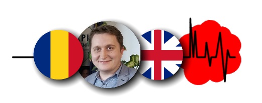
Dr Ruben Nechifor
Ruben will implement a new analysis approach for Dynamic Susceptibility Contrast MRI (DSC) in Quantiphyse, a free cross-platform analysis tool, for brain tumors, specifically addressing the corrections for leakage effects. Doing this, he will establish a DSC toolbox, optimized for brain tumor imaging, which can be used within the COST Action.
Affiliation: International Institute for Advanced Studies of Psychotherapy and Applied Mental Health (Cluj-Napoca)
Host: Institute of Biomedical Engineering, University of Oxford
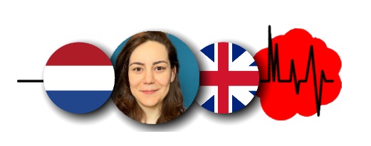
Ms Fatemehsadat Arzanforoosh
DSC MRI measurement of relative cerebral blood volume is the most commonly used approach for brain tumor perfusion imaging for predicting glioma grade, overall survival rate, and response to treatment. Fatemehsadat will improve the freely available analysis tool, Quantiphyse, to estimate tumor hemodynamic measurements in patient suffering from glioma.
Affiliation: Erasmus Medical Center (Rotterdam)
Host: Institute of Biomedical Engineering, University of Oxford
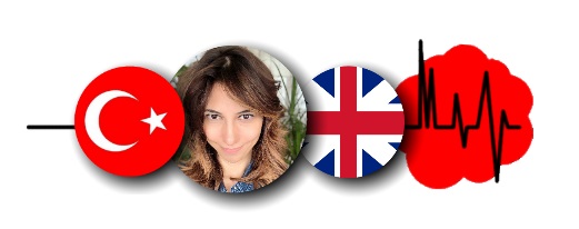
Dr Burcu Alparslan
Burcu will investigate if neuroimaging, performed at each component of the patient pathway after initial glioblastoma treatment, actually results in a real change in treatment management. She will mainly focus on DSC- and DCE-MRI.
Affiliation: Kocaeli University (Kocaeli)
Host: King’s College London Hospital (London)
Dr Golestan Karami
Golestan will connect knowledge with data during this multidisciplinary project. She will apply Deep Learning on a MRI perfusion dataset to predict the survival time of glioma patients.
Affiliation: Gabriele D’Annuzio University (Chieti)
Host: Leiden University
Mr Patrick Tang
Affiliation: Erasmus Medical Center (Rotterdam, the Netherlands)
Host: Aarhus University Hospital (Aarhus, Denmark)
Ms Yeva Prysiazhniuk
Yeva is investigating the clinical benefit of contrast (DSC) and no-contrast (ASL) perfusion MRI in diagnostics of glioma. The STSM established a start of the ongoing project in collaboration between Charles University and Oslo University Hospital, which focuses on the comparison of perfusion methods, their ability to differentiate tumor grades, and their predictive value in overall survival estimation. The aim of the project is to establish an optimal clinical approach for diagnostic brain tumor MR imaging
Affiliation: Charles University (Prague)
Host: Oslo University Hospital (Oslo)
Mr Hamail Ayaz
Hamail will investigate predominant recurrent tumours and radiation necrosis for a selected glioma patient cohort. This study will gather both conventional and advanced perfusion MRI data to automatically segment and compare the region of interest using deep learning algorithms for survival prediction.
Affiliation: Atlantic Technological University (Sligo)
Host: Adnan Menderes Üniversitesi (Aydin)
Mr Phillip Keil
Phillip investigated the use of independent component analysis of resting state functional magnetic resonance imaging data in the mapping of cortical areas involved in language in glioma patients. Usually, task based fMRI is employed to generate cortical maps for use in the planning of tumor resection surgery. However, Patients who already suffer heavy impairments in language due to tumor growth are at times unable to execute the necessary tasks during fMRI examination. If resting state fMRI, which does not require active contribution from the patient, can serve as an alternative, more patients who suffer tumor related functional impairment can benefit from fMRI assisted surgical planning.
Affiliation: University Hospital Cologne Center for Neurosurgery
(Cologne)
Host: Faculty of Sciences of the University of Lisbon (Lisbon)
STSM Awardees in COVID times
Ms Beatrice Cerqueti
During her STSM, Beatrice will learn and work on the segmentation of Diffuse Intrinsic Pontine Glioma, a pediatric tumor of the pons, on a dataset of MRI-images. This segmentation is a first step in defining normative tumor volume values, which is important for treatment administration.
Due to the current COVID-19 pandemic, this STSM is postponed until further notice.
Affiliation: University of Pavia (Pavia)
Host: University Medical Center (Amsterdam)
Ms Ayesha Mirchandani
Chemical Exchange Saturation Transfer MRI (CEST) is a new and promising biomarker for glioma prognosis and treatment. For a project in which CEST MRI will be compared with stereotactic biopsies, Ayesha will advance the translation from the CEST workflow, developed in the host institution, to her own institution and learn more about this technique.
Due to the current COVID-19 pandemic, this STSM is postponed until further notice.
Affiliation: King’s College London (London)
Host: Erasmus Medical Center (Rotterdam)
Ms Gökçe Hale Hatay
During her STSM, Gökçe will acquire knowledge about diffusion-weighted MRS and CEST methodologies: data acquisition, preprocessing and analysis. She will share this knowledge with her own research group.
Due to the current COVID-19 pandemic, this STSM is postponed until further notice.
Affiliation: Bogazici University (Istanbul)
Host: Leiden University Medical Center
Mr Umberto Villani
The Vascular, Extracellular, and Restricted Diffusion for Cytometry in Tumors (VERDICT) framework combines diffusion MRI data with physiological features inside tumors. Umberto will focus on the development of a physiology-driven formulation of this VERDICT model, specifically in gliomas.
Due to the current COVID-19 pandemic, this STSM is postponed until further notice.
Affiliation: Università degli studi di Padova (Padova)
Host: Medical Imaging and Visualization Centre (MMIV), Haukeland University Hospital
Ms Hande Halilibrahimoğlu
Hande will implement magnetic resonance spectroscopy, CEST, and diffusion-weighted MRS to create a machine-learning based multimodel MRS tool for glioma classification, thus enhancing diagnosis, grading and molecular subtyping of gliomas.
Due to the current COVID-19 pandemic, this STSM is postponed until further notice.
Affiliation: Bogazici University (Istanbul)
Host: Institut du Cerveau et de la Moëlle épinière (ICM) (Paris)
For further information regarding STSM’s, contact GliMR STSM coordinator, Ruben Nechifor at glimr.cost.applications@gmail.com.
